Microscopy at the Service of Food - 2nd Edition
Info utili


Sustainability: Drivers of Change
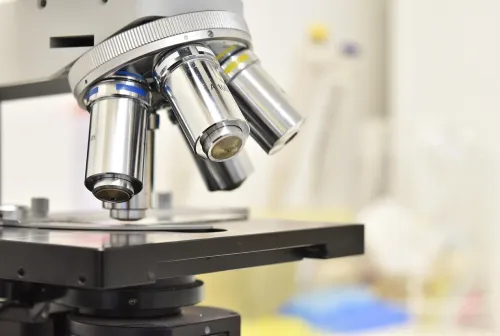
Microscopy has become a fundamental tool for studying the cellular effects of food components. This technique allows for in-depth analysis of cellular interactions and biochemical processes, offering valuable insights into how foods influence health and safety.
In vitro studies on the effects of foods
In vitro models are essential for analyzing the cellular responses triggered by different food components. Under controlled laboratory conditions, these models simulate real-world scenarios, allowing researchers to observe the morphological and functional changes of cells exposed to specific food elements. These studies are essential for understanding both the beneficial and adverse effects of foods on cellular behavior.
Preparation of Cytology Specimens
A crucial step in these studies is the careful preparation of cytology specimens. This process includes:
- Fixation and Permeabilization: Preservation of cellular structures while ensuring the penetration of staining reagents into the cells.
- Cytochemical/Immunocytochemical Reactions:These techniques allow the labeling of specific intracellular components, facilitating the visualization of target molecules and processes. This meticulous preparation is essential to obtain reliable and reproducible microscopic results.
Microscopic observation
Microscopic examination is performed using both brightfield microscopy and fluorescence microscopy:
- Brightfield microscopy: Provides an overall view of cell morphology, allowing for evaluation of cell structure and identification of any abnormalities.
- Fluorescence microscopy: Uses specific fluorochromes to highlight cellular markers of interest with high sensitivity and specificity. This technique allows for the detailed localization of proteins and other molecules within the cell.
Data Acquisition and Statistical Analysis
After microscopic observations, images are acquired using advanced imaging systems. They are then processed and analyzed with specialized software to extract quantitative and qualitative data. Statistical analysis is applied to validate the results, ensuring that the observed effects are significant and reproducible. This step is essential for correlating the in vitro effects of foods with potential biological implications.
The course was born from the need to stimulate the competitiveness of the food system in line with the "Smart" food theme, central to NODES Spoke 7. The training course is a strategic tool for developing highly specialized skills in the field of food microscopy. The module makes a significant contribution to promoting innovation and fostering a positive impact on the NODES area and the food sector.
The project is aimed at citizens, businesses, public administrations, researchers, and students who wish to expand their knowledge and acquire new skills in the use of in vitro models and microscopic techniques to study the effects of food.
Program
24/11/2025 | 10:00-12:00 The use of in vitro models in research (lecture): Dr. Ludovica Gaiaschi 14:00-16:00 The effects of natural compounds on cellular models (lecture): Prof. Maria Grazia Bottone, Dr. Federica Gola 4:00-6:00 PM Sterile room and preparation of in vitro preparations (lecture): instructors Dr. Fabrizio De Luca, Dr. Emma Lugli |
11/25/2025 | 9:00-12:00 Preparation of in vitro preparations (laboratory) 2:00-6:00 PM Extraction of biomolecules from natural compounds and in vitro treatment (laboratory) |
26/11/2025 | 10:00-12:00 Microscopy; From the invisible to the visible (lecture): Instructor Dr. Fabrizio De Luca 2:00 PM - 6:00 PM Preparation of cytological preparations for brightfield microscopy (laboratory) |
11/27/2025 | 9:00 AM - 12:00 PM Observation of cytological preparations under brightfield microscopy (laboratory) 2:00 PM - 6:00 PM Preparation of cytological preparations Cytological preparations for fluorescence microscopy (laboratory) |
28/11/2025 | 9:00-12:00 Observation of cytological preparations under fluorescence microscopy (laboratory) 2:00-6:00 PM Data acquisition and processing with statistical analysis concepts (mixed activity): instructors Dr. Fabrizio De Luca, Dr. Federica Gola, Dr. Ludovica Gaiaschi |
Lectures will be taught by professors, researchers, and doctoral students from the University of Pavia with proven knowledge of the topics covered.
The course will be held exclusively in person and will take place in classrooms and laboratories of the Department of Biology and Biotechnology "L. Spallanzani" of the University of Pavia. Locations will be communicated to participants promptly.
For information and questions, please send an email to the following addresses.
The following links contain (i) scientific papers recently published by our laboratory that report some of the techniques and models that will be used in the proposed training program and (ii) the laboratory's website:

Useful Information
Course Duration: 33 hours
Language: Italian
Delivery Method: In-person
Minimum Number of Participants: 1
Registration Fee: €1,500
